Introduction
Vision is arguably our most vital sense. It connects us to the world, allows us to navigate our daily lives, and holds the memories of the faces and places we love. Yet, because our sight often remains sharp for years, we tend to take it for granted. We treat our eyes as permanent, unchanging assets, failing to realize that many of the most serious eye conditions develop in complete silence.
For anyone concerned about their vision, or for those at higher risk due to age, diabetes, or family history, understanding what diagnostic technology is available is a powerful step toward maintaining independence. Trusted resources like BESTEYEHOSPITALS act as a bridge, helping patients identify centers of excellence that prioritize these diagnostic capabilities.
What Are Advanced Eye Diagnostic Services?
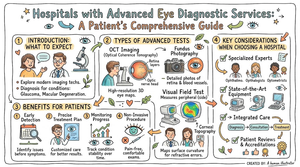
Advanced eye diagnostic services encompass a suite of specialized tests designed to examine the internal and external structures of the eye with extreme detail. Unlike a basic “refraction” or vision test that determines if you need glasses, diagnostic services focus on the health of the eye’s tissues.
The goal is structural and functional assessment. We are looking at the health of the optic nerve, the integrity of the retinal layers, the shape of the cornea, and the fluid dynamics within the eye. This data allows for personalized treatment planning; for instance, if we identify early-stage glaucoma, we can tailor the pressure-lowering medication specifically to the patient’s nerve damage patterns.
These services represent the intersection of high-speed computing, advanced optics, and medicine. They provide a “map” of the eye that allows ophthalmologists to track changes over time with clinical accuracy that was impossible just a few decades ago.
Why Early Eye Disease Detection Matters
The philosophy of modern eye care is simple: once vision is lost to disease, it is often impossible to fully restore. Therefore, our primary objective is to catch conditions before they ever impact your daily sight.
Early detection provides a wider window for therapeutic intervention. In the case of glaucoma, catching the condition early means we can initiate drops or laser treatments that stabilize the eye pressure, often preventing the permanent blindness that occurs in late-stage disease. For diabetic patients, retinal imaging can detect vascular leaks before they cause swelling in the macula.
Beyond the clinical benefits, early detection preserves quality of life. It allows patients to continue driving, working, and living independently. It reduces the need for invasive, last-resort surgeries and lowers the long-term financial and physical cost of managing advanced eye failure.
Table 1: Benefits of Advanced Eye Diagnostics
| Benefit | Patient Advantage |
| Early Detection | Allows for preventative therapy |
| Accurate Diagnosis | Avoids unnecessary, generic treatments |
| Vision Preservation | Keeps you independent for longer |
| Monitoring Progress | Provides objective proof of stability |
| Personalized Care | Treatment tailored to your unique eye anatomy |
Types of Advanced Eye Diagnostic Tests
The modern eye clinic is a hub of technological marvels. Here are the core tests that define high-level diagnostics.
Comprehensive Eye Examination
This is the baseline, involving an evaluation of the ocular surface, tear film, and internal structures after pupil dilation, providing a “big picture” view of eye health.
OCT (Optical Coherence Tomography)
This is arguably the most important advancement in recent decades. Think of it as an “optical biopsy.” It uses light waves to take cross-sectional pictures of your retina, allowing the doctor to see each individual layer of the nerve fiber.
Retinal Imaging
High-resolution fundus photography captures a detailed color map of your retina. This is essential for documenting the health of the blood vessels and the optic nerve, providing a baseline to compare against in future years.
Visual Field Testing
This measures your entire field of vision, including your peripheral sight. It is a critical tool for mapping the progress of glaucoma, as the disease typically steals peripheral vision first.
Corneal Topography
This test maps the curvature of the front surface of your eye (the cornea). It is essential for diagnosing conditions like keratoconus and is a mandatory step before any refractive surgery like LASIK.
Optical Biometry
Used primarily for cataract patients, this uses laser technology to measure the exact length of your eye. This is crucial for selecting the correct power for an intraocular lens during cataract surgery.
Eye Conditions Detected Through Advanced Diagnostics
Advanced testing allows us to see beyond the surface, detecting:
- Glaucoma: Detected through nerve fiber analysis (OCT) and peripheral vision testing.
- Cataracts: Evaluated through structural imaging and biometry to plan safe removal.
- Diabetic Retinopathy: Detected through retinal imaging that shows leaking vessels or micro-aneurysms.
- Age-Related Macular Degeneration (AMD): Identified via OCT, which shows thinning or fluid under the central retina.
- Retinal Detachment: Often preceded by “tears” that can be identified via high-resolution structural imaging.
- Keratoconus: Diagnosed when corneal topography shows an abnormal thinning and “coning” of the cornea.
Who Should Undergo Advanced Eye Testing?
Not everyone requires every test, but certain milestones and conditions warrant advanced care.
- Adults over 40: This is the baseline age where common age-related conditions like glaucoma begin to manifest.
- Diabetic Patients: Due to the risk of retinopathy, annual advanced retinal screening is non-negotiable.
- Family History: If your parents or siblings had glaucoma or macular degeneration, your baseline screening should start earlier.
- High-Risk Populations: Individuals with severe nearsightedness (myopia) are at higher risk for retinal detachments and should be monitored.
Table 3: Recommended Eye Screening Groups
| Patient Group | Recommended Evaluation |
| Adults 40+ | Baseline comprehensive exam |
| Diabetics | Annual retinal photography & OCT |
| Glaucoma Risk Patients | Periodic nerve fiber (OCT) and field testing |
| Cataract Patients | Surgical biometry and structural imaging |
| Patients with Vision Changes | Immediate advanced diagnostic testing |
How to Choose the Best Eye Diagnostic Hospital
When searching for hospitals with advanced eye diagnostic services, look for:
- Board-Certified Specialists: Your imaging is only as good as the ophthalmologist interpreting it.
- Technological Breadth: Does the center have an OCT? Do they have digital retinal mapping? A hospital that invests in a range of diagnostic tools is usually one that prioritizes evidence-based care.
- Integrated Care: The best diagnostic hospitals have a referral pipeline. If a test finds a problem, you want to be in a hospital where the surgeon or specialist is just down the hall.
- Transparency: They should provide you with copies of your scan data or explain the findings in plain, understandable language.
- Accreditation: Ensure the center meets the safety and quality standards required for specialized surgical or diagnostic work.
Preparing for an Eye Diagnostic Appointment
Eye tests are non-invasive and generally painless, but they do require preparation:
- Medical History: Bring a list of all your current medications, especially those for blood pressure or diabetes.
- Previous Records: If you have seen other specialists, bring your old test results or retinal scans.
- Dilation: Be prepared to have your pupils dilated. This makes the internal structures visible but will make you light-sensitive and blur your near vision for a few hours. Do not drive yourself to the appointment.
- Questions: Write down any symptoms, such as flashes, floaters, or blurriness, so you can describe them accurately.
Role of Technology in Modern Eye Care
We are living in the golden age of ophthalmology. AI-assisted diagnostics can now scan thousands of retinal images to detect the very first signs of diabetic retinopathy that a human eye might miss. Cloud-based records ensure that your diagnostic history follows you, allowing a doctor in a different clinic to see how your optic nerve has changed over the last five years. These tools don’t replace the doctor; they act as a super-powered assistant, ensuring that not a single detail is overlooked.
What Happens After an Eye Diagnosis?
An eye diagnosis is not just a label; it is the start of a plan. If a condition is detected, the specialist will stage it—grading the severity from early to advanced.
You might be placed on a “watch and wait” schedule, where you return for imaging every 6 months to monitor for changes. Or, you might be started on medication or recommended for a minor laser procedure. The goal is always to move you into a stabilized state where your vision is maintained at its current level for as long as possible.
Real-Life Patient Stories
- The Glaucoma Wake-Up Call: A 45-year-old accountant with no symptoms had a routine OCT scan. It revealed subtle nerve damage. By starting treatment that week, his specialist prevented the vision loss that would have otherwise occurred in his 50s.
- The Diabetic Milestone: A patient with long-term diabetes was monitored annually with retinal imaging. When small leaks were detected, laser treatment was applied in a single, painless session, preventing vision-threatening bleeding.
Future of Eye Diagnostics
The future is portable and personalized. We are developing smartphone-based retinal cameras that will allow eye screening in remote areas. We are also looking toward predictive analytics—using AI to predict your risk of vision loss five years in advance based on your current retinal architecture. The future of eye care is early, accessible, and incredibly accurate.
FAQs
- What are advanced eye diagnostic services?
Specialized, high-tech tests that map the eye’s structure. - What is an OCT scan?
An “optical biopsy” that creates 3D images of the retina. - How often should I get checked?
Every year after 40, or sooner if at risk. - Can diagnostics detect glaucoma early?
Yes, by measuring the optic nerve fiber layer. - Is retinal imaging safe?
Yes, it is a non-contact, light-based process. - What is corneal topography?
A map of the front of the eye. - Who needs these tests?
Diabetics, those with family history, and adults over 40. - Can diabetic eye disease be detected early?
Yes, via retinal imaging. - Are they painful?
No, they are non-contact and painless. - How long does it take?
Usually less than an hour. - How do I choose a hospital?
Look for advanced equipment and certified specialists. - What if disease is found?
A treatment plan is created to stabilize your vision. - Is it covered by insurance?
Most medically necessary diagnostic tests are. - Can eye diseases be prevented?
Many can be managed so they don’t cause blindness. - Why is early diagnosis important?
Because vision lost is often difficult to get back.
Final Conclusion
Your vision is the window through which you experience the beauty of the world. Caring for that window requires more than just picking out a stylish pair of glasses; it requires a commitment to monitoring the health of the intricate, delicate structures that make sight possible. Hospitals with advanced eye diagnostic services represent the pinnacle of this commitment.By integrating technologies like OCT, high-definition retinal imaging, and corneal mapping, these institutions have shifted the paradigm of eye care toward early detection and vision preservation. Do not wait for symptoms to appear. Whether it is managing a condition like diabetes or simply staying ahead of age-related changes, the technology exists today to give you peace of mind.