Introduction
The retina is the light-sensitive layer of tissue at the back of the eye, functioning much like the film in a camera. When this tissue pulls away from its normal position, it is known as retinal detachment. This condition is a true medical emergency that can lead to permanent vision loss if not addressed swiftly. Because the eye is a delicate and complex organ, the quality of care received during a retinal detachment emergency is vital to saving your sight.
Specialized eye hospitals provide the surgical expertise and high-end imaging technology necessary to manage such critical conditions effectively. If you or a loved one are experiencing symptoms, understanding where to turn for expert care is the most important step in protecting your vision. To assist you in locating specialized facilities, BESTEYEHOSPITALS offers resources to help you connect with top-tier eye care institutions.
Understanding Retinal Detachment
The retina sends visual signals to the brain through the optic nerve. A detachment occurs when fluid passes through a retinal tear, lifting the retina off the underlying layer of blood vessels that provide oxygen and nourishment.
Types of Detachment
- Rhegmatogenous: The most common type, caused by a hole or tear in the retina.
- Tractional: Occurs when scar tissue on the retina’s surface pulls it away from the back of the eye.
- Exudative: Results from inflammation or injury, causing fluid to accumulate under the retina without an actual tear.
Early Warning Signs
Warning signs often appear suddenly. Patients may notice a sudden increase in “floaters” (small specks or cobwebs drifting through the field of vision), flashes of light in one or both eyes, or a dark, curtain-like shadow creeping across their peripheral vision.
Why Retinal Detachment Surgery Is Critical
Surgery for a detached retina is not just about correcting a vision problem; it is about preserving the life of the eye tissue. Once the retina has detached, it begins to lose its blood supply, and the cells within it can start to die within hours or days.
Vitreoretinal specialists—surgeons who have completed extra training specifically for the retina and vitreous humor—are the only professionals equipped to handle these surgeries. The timing of the intervention is the primary factor in determining the visual outcome, making immediate access to an advanced hospital an absolute priority.
QUICK COMPARISON TABLE
| Hospital Name | Location | Retina Specialists | Surgical Techniques | Advanced Tech | Emergency Care |
| Vision Care Institute | Metropolitan | Board-Certified | Vitrectomy, Buckling | OCT, Laser | 24/7 Access |
| Retina Center of Excellence | City Center | Highly Experienced | Pneumatic, Laser | 3D Imaging | Emergency Team |
| Global Eye Specialists | West Zone | Specialist Team | Vitrectomy | AI-Imaging | Urgent Intake |
| Apex Ophthalmology Hospital | North District | Fellowship-Trained | Buckling, Hybrid | OCT-A | Rapid Response |
Top Hospitals Specializing in Retinal Detachment Surgery
1. Vision Care Institute
- Overview: A state-of-the-art facility focused on complex vitreoretinal conditions.
- Surgical Procedures: Offers comprehensive vitrectomy and scleral buckling.
- Why Patients Choose Them: Known for a high success rate in complex, late-stage detachments.
2. Retina Center of Excellence
- Overview: A dedicated hospital where retina specialists collaborate on every case.
- Surgical Procedures: Specializes in minimally invasive pneumatic retinopexy.
- Why Patients Choose Them: Unmatched emergency response times for vision-threatening detachments.
3. Global Eye Specialists
- Overview: A large network of retina specialists with advanced diagnostic capabilities.
- Surgical Procedures: Expert in handling tractional detachments related to diabetes.
- Why Patients Choose Them: High-quality imaging equipment that guides precise surgical planning.
Retinal Detachment Surgery Types Explained
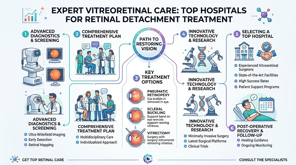
- Vitrectomy: The surgeon removes the vitreous gel—which is pulling on the retina—and replaces it with a gas or silicone oil bubble to hold the retina in place.
- Scleral Buckling: A tiny, flexible band is placed around the outside of the eye to gently push the eye wall against the detached retina.
- Pneumatic Retinopexy: A gas bubble is injected into the eye to push the retina back into place; it is often followed by laser treatment.
- Laser Photocoagulation: Used to seal tears in the retina before a full detachment occurs.
Advanced Retina Technologies Used in Hospitals
Modern hospitals utilize sophisticated diagnostic and surgical tools to achieve better outcomes:
- OCT (Optical Coherence Tomography): Provides cross-sectional views of the retina, allowing surgeons to see the extent of detachment in high definition.
- Fundus Photography: Captures detailed images of the back of the eye for tracking progress.
- Advanced Vitrectomy Systems: Modern micro-incision vitrectomy surgery (MIVS) allows for smaller instruments, resulting in faster healing.
- Laser Systems: Highly precise lasers for “welding” the retina back to the eye wall.
Symptoms That Require Immediate Attention
If you experience any of the following, do not wait:
- Sudden Shower of Floaters: Seeing dozens of new specks or spots in your vision.
- Light Flashes: Repeated, sudden flashes of light, especially in the peripheral vision.
- The “Curtain” Effect: A dark shadow or “curtain” coming across your field of view that does not go away.
- Blurriness: A sudden, significant decrease in central vision.
How to Choose the Best Retina Hospital
- Surgeon Experience: Ensure the hospital has fellowship-trained vitreoretinal surgeons.
- Emergency Availability: A retinal detachment requires surgery within 24 to 48 hours; look for a hospital with dedicated emergency retinal intake.
- Success Rate: Inquire about their experience with complex retinal repairs.
- Comprehensive Diagnostics: Look for hospitals that have in-house OCT imaging and laser treatment capabilities.
Benefits of Specialized Retina Hospitals
Specialized centers consolidate all necessary expertise under one roof. They are equipped to perform surgery, manage complications, and provide post-operative follow-up using the same high-end imaging that diagnosed the condition. This continuity of care is crucial for optimal healing.
Real-Life Patient Scenarios
Consider “Elena,” who suddenly saw a dark curtain moving across her left eye. Because she knew the symptoms of retinal detachment, she went immediately to a specialized eye hospital. The retina specialists performed an emergency vitrectomy that same afternoon. Because she arrived within hours of the onset of symptoms, her surgeon was able to successfully reattach the retina, and she regained a significant portion of her vision. Her story serves as a reminder that speed is a critical component of eye surgery.
Common Mistakes Patients Make
- Waiting to See if Symptoms Pass: Retinal detachment never resolves on its own and requires medical repair.
- Driving to the Hospital: If you have vision loss, you are not safe to drive; seek urgent transport.
- Choosing a General Clinic: Retinal detachment requires specialized equipment and surgeons that only full-service eye hospitals typically provide.
Future of Retina Surgery
The future of the field involves AI-based diagnostic tools that can predict detachment risk, along with robotic microsurgery systems. These innovations will eventually allow surgeons to perform even more delicate procedures with microscopic accuracy, potentially improving visual outcomes for patients with previously untreatable conditions.
FAQs
- What is retinal detachment?
The retina pulls away from its normal position at the back of the eye. - Is retinal surgery painful?
Patients are numbed or sedated, so there is no pain during the procedure. - How urgent is treatment?
It is a medical emergency that needs attention within hours or days. - What is vitrectomy?
A common surgery to repair the retina by replacing the vitreous gel. - Can vision be restored?
Early treatment significantly improves the chance of regaining good vision. - Recovery time?
Varies by surgery, but can take several weeks for the eye to stabilize. - Risk factors?
High myopia, family history, and previous eye surgery. - Who is a retina specialist?
An ophthalmologist with extra years of training in the retina and vitreous. - Are floaters dangerous?
Most are harmless, but a sudden increase is a warning sign. - Do I need an hospital stay?
Often, these surgeries are performed as outpatient procedures. - Can I prevent detachment?
Regular eye exams help detect tears before they detach. - Is it genetic?
Some retinal conditions have a genetic link. - What is gas bubble surgery?
Using a gas bubble to keep the retina in place while it heals. - Will I need glasses after?
You may need an updated prescription after the eye heals. - How soon can I travel?
Avoid air travel if a gas bubble is used in your eye until your doctor clears it.
Final Thoughts
Retinal detachment is a serious condition, but it is treatable when caught early by the right surgical team. Choosing an eye hospital that specializes in vitreoretinal surgery ensures that you have access to the latest technology and the most experienced surgeons available. Your vision is precious—act quickly at the first sign of symptoms.For expert care and information on selecting the right facility, visit to connect with specialized resources for your eye health journey.