Introduction
The ability to see the world around us with sharp clarity depends heavily on the frontmost layer of the eye known as the cornea. Acting as a protective window and a powerful focusing lens, this clear, dome-shaped surface bends light rays as they enter the eye, guiding them smoothly onto the retina at the back of the eye. However, when the cornea becomes scarred by severe infection, distorted by degenerative thinning, or swollen due to cellular loss, it turns cloudy and uneven. This structural degradation blocks or distorts light rays, causing severe visual impairment or even functional blindness that cannot be corrected with conventional glasses or standard contact lenses. For individuals facing advanced disease, undergoing surgical replacement remains the definitive medical path to restoring clear sight.
Navigating complex ophthalmic terminology and locating highly specialized surgical centers can feel overwhelming for patients. Dedicated medical platforms like BESTEYEHOSPITALS serve as a vital tool during this research phase. The platform helps patients explore eye hospitals, compare eye treatments, discover ophthalmology specialists, review corneal transplant programs, and make informed eye-care decisions. By selecting a high-volume, accredited clinical center equipped with advanced microscopic tools, you ensure that your delicate procedure is performed with the highest level of surgical safety.
Why Choosing the Right Hospital for Corneal Transplants Matters
A keratoplasty is an incredibly delicate, micro-surgical procedure that involves placing ultra-thin tissue layers under a specialized operating microscope. Unlike traditional open surgical procedures, cornea surgery requires managing tissue grafts that are sometimes only a few microns thick. Clinical outcome registries confirm that graft survival rates, endothelial cell preservation, and the long-term stabilization of vision are directly correlated with the annual procedural volume of the hospital and its operating medical team.
Furthermore, leading best eye hospitals look past basic community standards, housing advanced tracking infrastructure and specialized cleanroom environments. Donor cornea tissue must be carefully evaluated, screened, and stored in certified eye banks before it enters the operating room. Choosing a premier hospital ensures that you have access to specialized surgeons, cutting-edge surgical tools like femtosecond lasers, and emergency ophthalmic care if your body experiences an acute graft rejection episode.
Understanding the Cornea and Its Role in Vision
To understand why surgical replacement becomes necessary, it is helpful to look closely at the multi-layered structure of this delicate ocular tissue. The cornea does not use blood vessels to receive oxygen and nutrients; instead, it relies on tears on the outside and aqueous humor fluid on the inside to keep it clear and healthy.
       [Outside of Eye]
     ───────────────────────
      Layer 1: Epithelium     <── Protective barrier block
      Layer 2: Bowman's Layer <── Anchor membrane
      Layer 3: Stroma         <── Main structural thickness (90%)
      Layer 4: Descemet's     <── Deep protective wall
      Layer 5: Endothelium    <── Vital fluid pump keeping tissue clear
     ───────────────────────
       [Inside of Eye]
The Multilayered Structure
The cornea is comprised of five distinct, highly organized tissue layers:
- The Epithelium: The outermost, rapidly regenerating cellular layer that serves as a barrier against bacteria, foreign objects, and minor scratches.
- Bowman’s Layer: A smooth, dense sheet of acellular collagen fibers that anchors the epithelium securely to the structural layer below.
- The Stroma: The thickest layer, accounting for roughly $90\%$ of the cornea’s total thickness. It is composed of perfectly aligned, parallel collagen fibrils that provide structural strength while allowing light to pass through completely unobstructed.
- Descemet’s Membrane: A thin, highly elastic basement membrane that serves as a protective wall for the delicate innermost cell layer.
- The Endothelium: A vital, single layer of specialized cells lining the back surface of the cornea. These cells act as a continuous pump, drawing excess fluid out of the stroma to keep the cornea perfectly balanced and clear. Endothelial cells cannot multiply or regenerate; if they are destroyed by disease or trauma, the cornea loses its pumping power, absorbs fluid, and swells, causing permanent cloudiness.
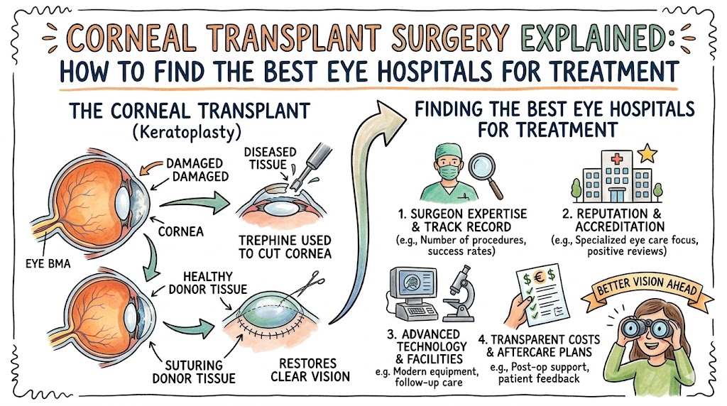
What Is a Corneal Transplant (Keratoplasty)?
When structural damage or cellular loss permanently compromises the clarity of your eye, a corneal transplant surgery becomes necessary. Clinically referred to as a keratoplasty, this procedure involves carefully removing the damaged, non-functional native tissue and replacing it with a healthy, clear cornea graft harvested from a deceased human donor.
The primary clinical goal of this intervention is to clear away structural distortions or cloudiness, instantly restoring a transparent pathway for light to travel into the eye. By using advanced microscopic stitching techniques or specialized gas bubbles to anchor the tissue, surgeons can remodel the eye’s shape, clear chronic fluid swelling, and restore crisp, functional vision to the patient.
Types of Corneal Transplant Procedures
Modern ophthalmology has moved away from old-fashioned approaches where the entire cornea was replaced for every condition. Today, specialized cornea specialists practice selective lamellar keratoplasty—replacing only the specific damaged layers of the eye while preserving your healthy, uninjured tissue layers intact.
                  ┌── Penetrating Keratoplasty (PK) ── Full-thickness replacement
                  ├── DALK Surgery ──────────────────── Front layers only (Stroma)
Keratoplasty ─────┼── DSEK Surgery ──────────────────── Endothelium + deep stroma
                  ├── DMEK Surgery ──────────────────── Pure single-cell layer pump
                  └── Keratoprosthesis ──────────────── Artificial plastic optic design
Penetrating Keratoplasty (PK)
A penetrating keratoplasty is a full-thickness transplant procedure. The surgeon uses a micro-blade or a high-precision femtosecond laser to cut out a circular button of the patient’s entire damaged cornea, replacing it with a full-thickness donor graft secured with ultra-fine nylon sutures. This approach is reserved for deep scars, advanced structural distortions, or injuries that impact all five layers of the tissue.
Deep Anterior Lamellar Keratoplasty (DALK)
DALK surgery is an advanced lamellar technique used when the inner endothelial pump is healthy, but the front structural layers (the stroma) are scarred or thinned out. The surgeon carefully peels away the damaged front layers down to Descemet’s membrane, leaving the patient’s native endothelial cells untouched. Because the inner layer is preserved, the risk of graft rejection is drastically lower than with a full-thickness transplant.
Descemet Stripping Endothelial Keratoplasty (DSEK)
DSEK surgery targets diseases that impact only the back endothelial pump layer. The surgeon removes the damaged inner cell layer along with a very thin piece of the structural stroma, replacing it with a partial-thickness donor graft. This tissue is inserted into the eye through a tiny incision and held in place using an internal air or gas bubble, eliminating the need for extensive surface stitches on the front of the eye.
Descemet Membrane Endothelial Keratoplasty (DMEK)
DMEK surgery represents the highest level of precision in endothelial transplantation. Instead of inserting a thick piece of stromal tissue like in DSEK, the surgeon transplants a pure, single-cell layer graft composed only of Descemet’s membrane and functioning endothelial cells. This ultra-thin tissue roll is carefully unfurled inside the eye and held in place using a gas bubble. Because it uses almost zero donor stromal tissue, it offers exceptionally fast vision recovery and a near-zero risk of long-term graft rejection.
Artificial Cornea (Keratoprosthesis)
When a patient has suffered severe chemical burns, multiple past graft failures, or advanced autoimmune conditions, their body may be unable to accept a natural human tissue transplant. In these complex cases, surgeons utilize an artificial cornea, known as a keratoprosthesis (such as the Boston KPro). This design pairs a clear plastic central optic lens with supportive donor tissue rings to restore a clear pathway for vision, even when traditional human grafts cannot survive.
Table 1: Types of Corneal Transplant Procedures
| Procedure Type | Primary Clinical Benefits | Average Vision Recovery Time | Ideal Candidate Profile |
| Penetrating Keratoplasty (PK) | Replaces all five layers; effectively clears deep scars or complex injuries. | $6 – 12$ months | Patients with full-thickness scars, severe perforations, or advanced multi-layer disease. |
| DALK Surgery | Near-zero risk of endothelial graft rejection; preserves natural inner cells. | $3 – 6$ months | Individuals with advanced keratoconus or front stromal scars with a healthy inner pump. |
| DSEK Surgery | Minimal surface stitches; faster healing; lower rejection risk than full PK. | $1 – 3$ months | Patients with Fuchs’ Dystrophy or corneal swelling with tricky inner anatomy. |
| DMEK Surgery | Exceptional visual clarity; ultra-fast recovery; lowest rejection risk among human grafts. | $2 – 6$ weeks | Individuals with isolated endothelial failure or Fuchs’ Dystrophy needing rapid visual recovery. |
| Keratoprosthesis (KPro) | Does not require a living human graft; works in high-risk eyes prone to rejection. | $1 – 4$ weeks | Patients with multiple past graft failures, severe chemical burns, or ocular pemphigoid. |
Signs You May Need a Corneal Transplant
Corneal diseases often develop slowly, with early symptoms masquerading as simple prescription changes. However, as the tissue continues to degrade or thin, clear warning signs appear, signaling the need for an evaluation at an advanced center for corneal disease treatment.
- Progressive, Uncorrected Blurred Vision: Visual clarity decreases steadily over time, and regular adjustments to glasses or specialty hard contact lenses no longer improve your vision.
- Severe Monocular Diplopia: Experiencing ghost images, severe glare, or double vision out of a single eye, caused by irregular astigmatism as the cornea thins and warps.
- Extreme Photophobia and Ocular Pain: Light sensitivity becomes so intense that being in normal room lighting causes discomfort or throbbing pain, which often indicates deep tissue swelling or raw epithelial blisters (bullae) on the surface of the eye.
- Visible Cloudiness or Whitening: The normally clear, invisible front window of the eye takes on a hazy, milky, or dense white appearance that can be seen in a mirror.
- Recurrent Ocular Surface Irritation: Experiencing frequent bouts of tearing, a foreign body sensation, or shooting pains as small fluid blisters on a swollen cornea break open on the surface of the eye.
Common Causes of Corneal Damage and Disease
Anatomical damage to the cornea can trace its roots to genetic conditions, chronic eye rubbing, deep infections, or surgical changes over time.
- Fuchs’ Endothelial Dystrophy: A progressive, hereditary condition where endothelial cells die off faster than normal. As the cell count drops, the inner pump fails, causing fluid to accumulate in the stroma and cloud vision.
- Keratoconus: A degenerative thinning disorder where the structural stroma weakens, causing the cornea to bulge outward into an irregular, cone-like shape that causes severe distortion.
- Pseudophakic Bullous Keratopathy (PBK): Chronic tissue swelling that can develop after cataract surgery if the endothelial cells sustain unavoidable stress during the procedure.
- Infective Corneal Ulcers: Deep, destructive scars left behind by severe bacterial, fungal, or viral infections (such as Herpes Simplex Keratitis or Acanthamoeba from contact lens misuse).
- Mechanical Eye Injuries: Direct cuts, chemical splashes, or flying debris that tear the structural layers of the eye, leaving behind thick, permanent scar tissue.
Table 3: Common Corneal Diseases and Treatment Options
| Condition | Primary Clinical Symptoms | Recommended Treatment Options |
| Fuchs’ Dystrophy | Hazy morning vision that clears slowly; severe glare; painful surface blisters. | Partial-thickness endothelial transplant (DMEK or DSEK surgery). |
| Advanced Keratoconus | Progressive astigmatism; ghost imaging; severe structural thinning. | Custom scleral contact lenses, corneal cross-linking (early), or DALK surgery. |
| Corneal Scarring | Fixed, cloudy white spot over the pupil; permanent loss of clarity. | Penetrating Keratoplasty (PK) or DALK if the inner layer is unaffected. |
| Bullous Keratopathy | Chronic water swelling; low visual acuity; persistent pain from surface blisters. | Specialized hypertonic saline drops (early) or endothelial tissue replacement. |
What Makes the Best Hospitals for Corneal Transplants Stand Out?
When searching for the best hospitals for corneal transplants, look past basic healthcare marketing. Premier eye centers are distinguished by several key quality standards and institutional capabilities:
Elite Cornea Fellowship-Trained Surgeons
Top centers recruit ophthalmologists who have completed intensive, dedicated fellowship training focusing exclusively on cornea and external disease. These specialists maintain high annual case volumes, which has been clinically proven to lower complication rates and improve long-term graft survival.
Integration with Certified Eye Banks
Leading corneal transplant hospitals maintain direct partnerships with accredited eye banks. This ensures that donor corneas are harvested, evaluated using specular microscopy to verify healthy endothelial cell counts, and delivered to the operating room under strict sterile storage controls.
Advanced Diagnostic and Micro-Surgical Infrastructure
The best hospitals utilize high-resolution anterior segment optical coherence tomography (AS-OCT) and digital specular microscopes to track your tissue thickness down to the micron. Their operating rooms feature advanced 3D heads-up visualization systems, high-precision femtosecond lasers for cutting perfect graft edges, and active intraoperative neuromonitoring tools to ensure the highest safety.
Advanced Technologies Used in Modern Corneal Transplant Surgery
Ophthalmic surgery changes rapidly, combining micromanipulation tools with digital guidance systems to maximize visual outcomes.
- Femtosecond Laser-Assisted Keratoplasty (FLAK): Instead of using mechanical hand-blades, surgeons can use an ultra-fast laser to cut matching zig-zag or top-hat shapes into both the patient’s eye and the donor tissue, creating a perfect fit that heals faster with less astigmatism.
- 3D Heads-Up Surgical Visualization: Surgeons wear specialized polarized glasses and look at a large, high-definition 4K 3D monitor rather than leaning over standard microscope eyepieces. This provides unparalleled depth perception and clarity when handling delicate, micron-thin tissue grafts.
- Intraoperative OCT Tracking: High-resolution scans are captured in real time during the procedure, allowing the surgeon to confirm that an ultra-thin DMEK graft is facing the correct direction and perfectly flat against the back of the eye before finishing the surgery.
Benefits of Choosing Leading Corneal Transplant Centers
Entrusting your vision recovery to an established, top-tier eye hospital offers several key advantages:
- Lower Rates of Graft Rejection: Precision graft matching and advanced medications reduce the risk of your immune system attacking the donor tissue.
- Superior Visual Clarity: Advanced laser cuts and precision stitching techniques minimize postoperative astigmatism, helping you achieve sharper vision.
- Reduced Risk of Infection: Operating out of dedicated ophthalmic suites helps minimize the risk of endophthalmitis or deep eye infections.
- Comprehensive Long-Term Management: Integrated follow-up pathways ensure that stitches are removed safely and your eye health is tracked carefully over time.
Risks and Complications of Corneal Transplant Surgery
While modern techniques have made a keratoplasty highly reliable, it is a significant intraocular intervention that carries specific risks that require careful consideration:
- Acute Immunological Graft Rejection: Your immune system can recognize the donor cornea as foreign tissue at any time. If rejection occurs, it causes sudden swelling and cloudiness, which requires immediate treatment with intensive steroid eye drops to save the graft.
- Postoperative Graft Astigmatism: Because human tissue requires stitches or gas bubbles to heal, the new cornea can settle unevenly, causing astigmatism that may require specialized custom contact lenses or minor laser reshaping later on.
- Secondary Glaucoma: The frequent use of steroid eye drops after surgery can cause intraocular pressure to rise in some patients, requiring careful monitoring and pressure-lowering drops to protect the optic nerve.
- Graft Failure or Decompensation: Over time, the endothelial cells on a donor graft can slowly decrease in number. If the cell count drops too low, the graft can become cloudy, requiring a simple partial-thickness replacement.
- Intraocular Infection (Endophthalmitis): A rare but serious complication where bacteria or fungi enter the eye during or after surgery, requiring urgent treatment with intraocular antibiotics.
Factors Patients Should Consider Before Choosing a Hospital
Selecting the right eye hospital requires checking objective quality metrics, facility accreditations, and long-term follow-up care options against your personal health needs.
Table 2: Factors to Consider Before Choosing an Eye Hospital
| Selection Factor | Clinical Importance | Essential Questions to Ask the Hospital |
| Surgeon Volume & Background | High annual volumes ensure the surgeon is skilled in delicate lamellar techniques like DMEK. | “Is my surgeon cornea-fellowship trained, and how many DMEK/DALK procedures do they perform annually?” |
| On-Site Eye Bank Access | Ensures donor tissue is screened thoroughly and verified for healthy cell counts. | “Does the facility partner with an accredited eye bank to verify donor tissue endothelial cell counts?” |
| Emergency Availability | Graft rejection requires immediate treatment within 24 hours to prevent permanent tissue failure. | “Do you have a dedicated cornea specialist available 24/7 to manage acute graft rejection emergencies?” |
| Advanced Micro-Imaging Tools | Intraoperative OCT and 3D imaging are essential for verifying proper graft placement. | “Does your operating room feature intraoperative OCT imaging for layer verification?” |
Questions Patients Should Ask Their Eye Surgeon
Before consenting to surgery, schedule a detailed consultation to review your diagnostic scans and treatment options. Use this practical checklist to help guide your discussion:
- “Based on my diagnostic scans, am I a candidate for a partial-thickness transplant (DMEK/DALK) or do I need a full-thickness PK?”
- “What is the average endothelial cell count of the donor tissue you select for your patients?”
- “How many stitches will be placed on my eye, and when do you anticipate they will be safely removed?”
- “What is your personal success rate and graft survival rate for this specific type of cornea surgery?”
- “How long must I use steroid eye drops after surgery, and how will we monitor my intraocular pressure?”
- “When can I safely return to low-impact daily activities, driving, and returning to my job?”
Cost Factors Associated With Corneal Transplants
The total financial investment required for cornea replacement varies based on the type of procedure, facility infrastructure, and insurance coverage structures.
Insurance and Tissue Fee Dynamics
Because a corneal transplant is a medically necessary, sight-restoring procedure, it is widely covered by Medicare and major commercial health insurance providers. However, out-of-pocket expenses can vary based on deductibles and whether your providers are in-network. A unique factor in ophthalmic surgery is the cornea tissue acquisition fee, which covers the cost of harvesting, testing, and storing the donor tissue at the eye bank—an expense that must be pre-authorized with your insurance team.
Direct and Indirect Costs
- Direct Expenses: Pre-operative specular microscopy, corneal topography, surgical suite fees, donor tissue acquisition fees, surgeon and anesthesiologist fees, and prescription steroid eye drops.
- Indirect Expenses: Transportation to frequent follow-up appointments, protective shields, time away from work during early recovery, and custom scleral contact lenses needed later to correct astigmatism.
Recovery Timeline After Corneal Transplant Surgery
Recovering from cornea surgery is a slow, gradual process that requires patience and a commitment to protecting your healing eye from physical stress or injury.
Table 4: Recovery Timeline After Corneal Transplant Surgery
| Recovery Stage | What to Expect Post-Operative | Recommended Care & Patient Guidelines |
| Days 1–7 | Mild irritation or a scratchy feeling; vision may be blurry; partial-thickness patients must maintain a flat, face-up position. | Use your prescribed antibiotic and steroid drops exactly as directed; wear your protective clear eye shield 24/7; avoid rubbing your eye. |
| Weeks 2–4 | Swelling begins to clear; visual clarity improves gradually; discomfort decreases. | Continue your regular steroid drop schedule; protect your eye from water during showers; avoid lifting anything over 10 pounds. |
| Months 2–6 | Vision stabilizes but may fluctuate as stitches adjust; intraocular pressure is tracked closely. | Attend all follow-up appointments; wear protective eyewear outdoors; do not engage in contact sports or swimming. |
| Months 6–12+ | Surface stitches are systematically removed or adjusted; final vision correction options are explored. | Transition to a low-dose long-term drop schedule; get fitted for glasses or custom scleral contact lenses to optimize clarity. |
Post-Surgery Eye Care and Long-Term Vision Management
Achieving your best final vision depends heavily on the daily habits and care routines you maintain at home after leaving the surgical center.
Proper Eye Drop Technique
Steroid eye drops are essential for preventing graft rejection. Wash your hands thoroughly before applying drops, pull down your lower eyelid slightly to create a small pocket, and close your eye gently for one minute without blinking or squeezing to let the medication absorb fully.
Protecting Your Healing Eye
Never rub your eye under any circumstances. Rubbing can break fine surface stitches, cause a gas bubble to shift out of place, or displace a delicate tissue graft. Wear your clear plastic eye shield every night while sleeping for at least the first 4 to 6 weeks to prevent accidental rubbing.
Lifestyle Tips for Protecting Corneal Health
To support long-term healing and protect your vision investment, integrate these eye-healthy practices into your daily routine:
- Wear UV-Blocking Sunglasses: Protect your healing graft from ultraviolet rays by wearing wrap-around sunglasses outdoors, which also helps minimize glare and light sensitivity.
- Avoid Smoky or Dusty Environments: Stay away from environments with heavy smoke, chemical fumes, or blowing dust, as these particles can irritate the ocular surface and increase your risk of infection.
- Practice Good Eye Hygiene: Wash your hands frequently, keep your eyelids clean, and never use expired eye drops or touch the tip of the drop bottle directly to your eye.
- Stay Well Hydrated: Drink plenty of water throughout the day and use preservative-free artificial tears if recommended by your doctor to keep your corneal surface smooth and comfortable.
Common Myths vs. Facts About Corneal Transplants
- Myth: An eye transplant involves removing the entire eyeball from a donor and connecting it to the patient’s nerves.
- Fact: It is impossible to transplant a whole eyeball because the optic nerve cannot be reconnected. A corneal transplant replaces only the clear front window of the eye, leaving the rest of your eye structure completely intact.
- Myth: Corneal transplant surgery is incredibly painful with a very difficult early recovery.
- Fact: The procedure is performed under local numbing blocks, making it painless. Most patients experience only a mild scratchy feeling or dull ache during early recovery, which is easily managed with over-the-counter pain relievers.
- Myth: Your body can never reject a corneal transplant once the first year of healing has passed.
- Fact: Ocular graft rejection can happen at any time, even decades after a successful procedure. Patients must know the warning signs and keep steroid drops on hand throughout their life.
- Myth: You will achieve perfect, 20/20 vision right after your cornea surgery is completed.
- Fact: Healing is a slow process. Vision improves gradually over several weeks or months, and most patients will need glasses or custom contact lenses to achieve their sharpest final vision.
Latest Innovations in Cornea Surgery and Eye Care
Ophthalmic science continues to advance, introducing innovative techniques designed to simplify care and improve patient outcomes.
Descemet Membrane Endothelial Transfer (DMET)
For patients with early-stage endothelial failure, researchers are studying an innovative technique where a donor endothelial graft is placed inside the eye without removing the patient’s native tissue. The donor cells naturally migrate to where they are needed, helping to clear fluid swelling without a complex tissue swap.
Non-Surgical Corneal Cross-Linking (CXL)
For early-stage keratoconus, specialists utilize corneal cross-linking (CXL) to stop disease progression before a transplant is ever needed. This in-office procedure pairs specialized riboflavin (Vitamin B2) drops with controlled UV light to strengthen the collagen bonds within the stroma, stabilizing the cornea’s shape and preserving vision.
Future of Corneal Transplant Technology
Looking ahead, eye care is moving toward bio-engineered and non-tissue solutions to help address global donor tissue shortages.
3D Bio-Printed Synthetic Stroma Grafts
Scientists are developing advanced 3D bio-printing techniques that use biocompatible collagen hydrogels to construct custom structural layers. These synthetic grafts can be tailored to match a patient’s exact corneal thickness and shape, which may eventually eliminate the need for human donor stroma tissue.
Endothelial Cell Culturing and Injections
An exciting advancement in vision restoration involves cell culturing. Instead of transplanting a full tissue layer from a donor, scientists can harvest a small number of healthy endothelial cells and multiply them millions of times in a lab. These cultured cells can then be injected directly into a patient’s eye through a tiny needle, where they settle and restore the inner pump without a traditional surgical transplant.
Expert Recommendations
Clinical guidance from leading international ophthalmology societies highlights the importance of patient awareness and specialized care:
Clinical Consensus: “Modern cornea surgery focuses on tissue preservation and micron-level precision (Zancanaro, 2026). Moving away from traditional full-thickness transplants toward selective layer surgeries like DMEK has dramatically improved visual outcomes and reduced rejection risks. However, the long-term success of these advanced procedures relies heavily on proactive patient care. Patients must understand their post-operative care routines, maintain a strict eye drop schedule, and seek care immediately if they notice any sudden drops in vision or redness, ensuring their graft remains healthy and clear for life.”
Common Mistakes Patients Should Avoid
Achieving your best final vision relies on making well-informed choices before surgery and practicing excellent judgment during recovery.
- Neglecting Your Eye Drop Schedule: Skipping doses of your prescribed steroid eye drops can weaken your eye’s protection, allowing your immune system to attack the donor tissue and potentially causing graft failure.
- Rubbing Your Eye During Early Healing: Rubbing your eye can shift a delicate graft out of place, disrupt healing tissue layers, or break micro-stitches, which can cause significant setbacks in your recovery.
- Getting Tap Water in Your Eye Too Soon: Avoid swimming or letting tap water run directly into your face during showers during the first few weeks, as water can carry harmful pathogens like Acanthamoeba into a healing incision.
- Ignoring Sudden Vision Changes: Never wait to see if symptoms like a sudden drop in vision, redness, or pain will improve on their own. These are early warning signs of graft rejection that require immediate evaluation within 24 hours to protect your sight.
Patient Safety Checklist
Proper preparation before arriving at the eye hospital helps ensure your procedure goes smoothly and sets a strong foundation for recovery.
Pre-Operative Preparation
- Verify Surgeon Fellowships: Confirm that your operating ophthalmologist has completed a formal, accredited fellowship in cornea and external disease.
- Arrange Post-Op Transport: Secure a trusted friend or family member to drive you home after surgery, as your vision will be blurred and you cannot drive safely.
- Set Up Your Recovery Space: Prepare a comfortable resting area at home with plenty of pillows so you can rest flat on your back, face-up, if your procedure requires a gas bubble.
- Clear Active Infections: Ensure your eyelids and surrounding skin are completely clear of active blepharitis, styes, or facial skin infections before surgery.
Post-Operative Monitoring
- Keep Emergency Contacts Handy: Place your cornea specialist’s 24/7 emergency contact number in an easily accessible spot on your phone or refrigerator.
- Wear Your Eye Shield Consistently: Keep your clear plastic eye shield securely taped over your eye whenever you sleep to prevent accidental rubbing.
- Track Your Drop Routine: Use a simple logbook or smartphone reminder app to ensure you take your antibiotic and steroid drops exactly as scheduled.
- Watch for Rejection Signs: Remember the RSVP protocol: contact your surgeon immediately if you experience Redness, Severe pain, a drop in Vision, or light sensitivity (Photophobia).
Key Takeaways
- Targeted Layer Surgery: Modern cornea replacement focuses on selective layer surgery (like DMEK or DALK), replacing only the damaged tissue while preserving your healthy layers.
- Prioritize Surgeon Expertise: Always select a cornea-fellowship trained specialist operating within a high-volume, accredited eye care hospitals network.
- The Rejection Warning Protocol: Remember the RSVP checklist: Redness, Severe pain, a drop in Vision, or Photophobia require immediate medical care within 24 hours.
- Commit to Home Care: Protecting your sight requires strict adherence to your steroid drop schedule, keeping your eye protected with a clear shield, and never rubbing your eye.
- A Gradual Sight Recovery: Vision improves slowly over weeks or months, and most patients will need glasses or custom scleral lenses to achieve their sharpest final vision.
Frequently Asked Questions
1. How long does a corneal transplant surgery take?
A standard partial-thickness transplant (like DMEK or DSEK) typically takes between 45 and 60 minutes. A full-thickness penetrating keratoplasty is slightly longer, usually taking 60 to 90 minutes depending on your eye anatomy.
2. Can I reject a corneal transplant years after the surgery?
Yes. Ocular graft rejection can happen at any time, even decades after a successful procedure. Because your immune system can always recognize donor tissue as foreign, maintaining your follow-up routine and using your drops as directed is essential.
3. What does the RSVP protocol mean for cornea patients?
The RSVP protocol stands for Redness, Severe pain, a drop in Vision, and light sensitivity (Photophobia). If you notice any of these warning signs, contact your cornea specialist immediately, as they can flag an early graft rejection episode.
4. Why must some partial-thickness patients lie flat on their backs after surgery?
Endothelial transplants like DMEK or DSEK use an internal gas bubble to hold the thin donor tissue up against the back of your eye. Lying flat on your back, face-up, ensures that buoyancy pushes the bubble in the right direction, sealing the graft perfectly.
5. Are donor corneas matched for blood type or eye color?
No. Because the cornea is naturally avascular (contains no blood vessels), it does not require blood type matching or HLA tissue typing for standard cases. Eye color is determined by the iris, which remains untouched during surgery.
6. When can I safely return to driving after surgery?
Your driving timeline depends on the visual acuity of your other eye and how quickly your transplant heals. Some endothelial transplant patients can drive within a few weeks, while full-thickness procedures can take several months before vision stabilizes enough for safe driving.
7. How long do surface stitches stay in the eye after a full-thickness transplant?
Sutures used in a full-thickness transplant (PK) heal very slowly. They are typically left in place for 6 to 12 months, and your surgeon will systematically remove or adjust individual stitches over time to manage astigmatism.
8. What is the difference between DSEK and DMEK surgery?
DSEK transplants the donor endothelial cell layer along with a very thin supportive piece of stromal tissue. DMEK transplants a pure, ultra-thin graft composed only of Descemet’s membrane and functioning endothelial cells, offering faster vision recovery and a lower rejection risk.
9. Can I get a corneal transplant if I have advanced glaucoma?
Yes, you can undergo a corneal transplant if you have glaucoma, but your intraocular pressure must be carefully managed beforehand. Your care team will monitor your eye pressure closely after surgery, as steroid drops can sometimes cause pressure to rise.
10. Where do donor corneas come from?
Donor corneas are harvested from deceased individuals who chose to donate their tissues. The tissue is managed by certified eye banks, where it undergoes extensive testing to ensure it is clear, healthy, and completely safe for transplantation.
11. Can a corneal transplant correct severe nearsightedness or farsightedness?
The primary goal of a transplant is to clear cloudiness and restore tissue clarity, not to correct refractive errors. While the procedure alters your eye’s shape, most patients will still need glasses or custom scleral contact lenses to achieve sharp focus.
12. What are scleral contact lenses, and why are they used after surgery?
Scleral lenses are large-diameter, hard contact lenses that vault completely over the cornea, resting on the white part of the eye (the sclera). They create a smooth, fluid-filled surface over an irregular or astigmatic cornea, providing exceptional visual clarity after surgery.
13. Is general anesthesia required for cornea surgery?
Most corneal transplants are performed using local numbing blocks paired with mild intravenous sedation. This keeps you relaxed, comfortable, and pain-free throughout the procedure without the longer recovery time of general anesthesia.
14. What happens if a corneal transplant fails completely?
If a graft fails over time due to natural cell loss or severe rejection, the procedure can be safely repeated. A surgeon can perform a second transplant—often using a quick, partial-thickness endothelial layer swap—to restore clarity to the eye.
15. How can I find out an eye hospital’s specific success rates for cornea surgeries?
You can confirm a facility’s credentials and performance data by reviewing public registry databases provided by national ophthalmology boards or checking listings on trusted platforms like [suspicious link removed], which ensure the center uses certified specialists and advanced diagnostic tools.
Conclusion
Best Hospitals for Corneal Transplants are defined by their commitment to clinical excellence, specialized surgeon choices, and advanced microscopic imaging infrastructure. Replacing a damaged or cloudy cornea using modern, selective layer surgery offers a reliable path to restoring clear vision and improving your quality of life.Rather than settling for basic care, prioritize facilities that partner with certified eye banks and offer specialized long-term follow-up care. Utilizing trusted research networks like helps patients discover leading eye hospitals, compare ophthalmology services, explore cornea specialists, review eye surgery options, and access information about advanced eye-care treatments worldwide. The platform highlights corneal transplantation, cataract surgery, LASIK, glaucoma care, retinal treatment, and global eye-care destinations. By working closely with an experienced cornea specialist, maintaining your eye drop routine, and protecting your eye during recovery, you can safeguard your vision investment and enjoy a bright, clear future.