Corneal transplantation, also known as corneal grafting or penetrating keratoplasty, is a surgical procedure where a damaged or diseased cornea is replaced by donated corneal tissue. The cornea is the clear part of eye in front of the iris and pupil. An unhealthy cornea affects your vision by scattering or distorting light and causing blurry or glary vision. A cornea transplant may be necessary to restore your functional vision. The surgical procedure is performed by ophthalmologists, medical doctors who specialize in eyes, and are often done on an outpatient basis. When corneal transplant surgery is indicated, the patient is registered with the Eye Bank. It is performed using general anesthesia or local anesthesia plus IV sedation.
A cornea transplant may be suggested due to any of the following reasons:
Scarring from infections, such as eye herpes or fungal keratitis.
Eye diseases such as keratoconus.
Hereditary factors or corneal failure from previous surgeries.
Thinning of the cornea and irregular shape (such as with keratoconus).
Complications from LASIK.
Chemical burns on the cornea or damage from an eye injury.
Excessive swelling (edema) on the cornea.
Procedure:
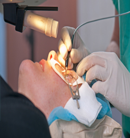
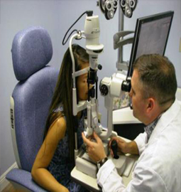
On the day of the surgery, the patient is given a brief physical examination by the surgical team and is taken to the operating room in the hospital or an outpatient surgery center. A local or general anesthesia may be used, depending on the health, age of the patient. and whether or not you prefer to be asleep during the procedure. With local anesthesia, an injection into the skin around your eye is used to relax muscles that control blinking and movement, and eye drops are used to numb the eye itself.
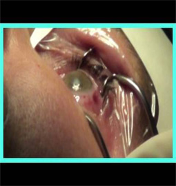
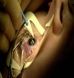
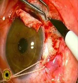
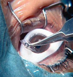
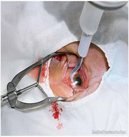
An eyelid speculum is used to keep the lids open, along with suitable lubrication to prevent the eye from drying. A metal ring is then stitched to the sclera, which will provide a base for a trephine. The surgeon inspects and measures the affected corneal area in order to determine the size of the transplantation. A trephine is then placed over the cornea and is used by the surgeon to cut the host cornea. The trephine is then removed and the surgeon cuts a circular graft from the donor cornea. A tissue nearly identical in shape is then sutured into place. Once this is done, the surgeon returns to the patient's eye and removes the host cornea.
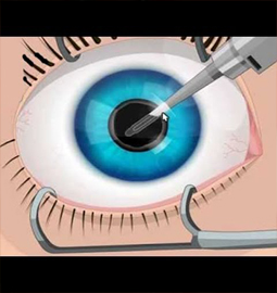
The donor cornea is then brought into the surgical field and maneuvered into place with forceps. Once in place, the surgeon will fasten the cornea to the eye with a running stitch (as used in the upper image above) or a multiple interrupted stitches (as in the lower image). The surgeon finishes up by reforming the anterior chamber with a sterile solution injected by a cannula, then testing that it's watertight by placing a dye on the wound exterior. With the metal ring removed and antibiotic eyedrops placed, the eye is patched, and the patient is taken to a recovery area while the effects of the anesthesia wear off. The patient typically goes home following this and sees the doctor the following day for the first post operative appointment. A plastic shield over your eye to protect it from being inadvertently rubbed or bumped. The procedure takes approximately two hours.
Risks associated with it:
While the cornea is avascular, there is still a potential for some blood loss, usually from suturing the metal ring to the sclera. Any blood loss is typically less than 2ml.
There is also a risk of infection. Since the cornea has no blood vessels (it takes its nutrients from the aqueous humor) it heals much slower than a cut on the skin. While the wound is healing, it is possible that it might become infected by various microorganisms. This risk is minimized by antibiotic prophylaxis (using antibiotic eyedrops, even when no infection exists).
Graft failure can occur at any time after the cornea has been transplanted, even years or decades later. The causes can vary, though it is usually due to new injury or illness. Treatment can be either medical or surgical, depending on the individual case. An early, technical cause of failure may be an overly tight stitch cheesewiring through the sclera.
Recovery tips:
For the first several weeks, heavy exercise and lifting are prohibited. However, you should be able to return to work three to seven days after surgery, depending on your job. Steroid eye drops will be prescribed for several months to help your body accept the new corneal graft. You should keep your eye protected at all times by wearing a shield or a pair of eyeglasses so that nothing inadvertently bumps or enters your eye. Stitches may be removed three to 17 months post-surgery, depending on the health of your eye and the rate of healing. Adjustments may be made to the sutures surrounding the new cornea to help reduce the amount of astigmatism resulting from an irregular eye surface.
About Eye
Eye Diseases
Vision problems
Find Cost
Surgeries & Treatments
Best Eye Hospitals
Testimonials
Get Free Quotes
Cataract Surgery
Intacs
Astigmatic Keratotomy(AK)
Canalicular Tear Repair
Chalazion Surgery
Conductive Keratoplasty(CK)
Corneal Transplantation
Lasik Surgery
Top Hospitals In India
Top Hospitals In Turkey
Top Hospitals In Mexico
Top Hospitals In Costa Rica
Top Hospitals In Singapore
Top Hospitals In UAE
Top Hospitals In Australia
Top Hospitals In Malaysia
Top Hospitals In Thailand
Top Hospitals In Phillipines