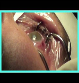


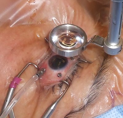
Scleral buckling surgery is the most common way to treat a retinal detachment.In this procedure a piece of silicone or sponge is sewn onto the sclera at the site of a retinal tear which pushes the sclera toward the retinal tear. This buckle supports the retina against the sclera till the scarring seals the tear.
Procedure:
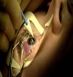
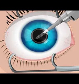
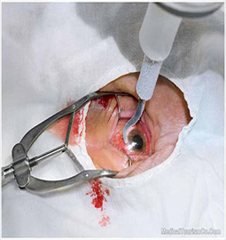
A scleral buckle is a piece of silicone sponge, rubber, or semi-hard plastic that is sewn into place on the outside of the eye. The buckling element is usually left in place permanently. This pushes in, or "buckles," the sclera toward the middle of the eye thus relieving the pull on the retina, allowing the retinal tear to settle against the wall of the eye. The buckle effect may cover only the area behind the detachment, or it may encircle the eyeball like a ring. This does not prevent a retinal break from opening again. Cryopexy, diathermy or laser photocoagulation is used to scar the retina and hold it in place until a seal forms between the retina and the layer beneath. The seal holds the layers of the eye together thus preventing fluid from getting between them. A gas bubble can also be injected into your eye to flatten the retina.
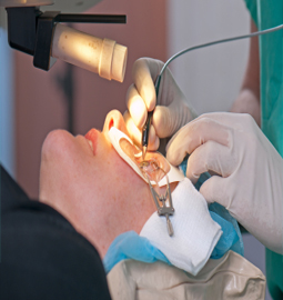
This procedure is done in a hospital as an outpatient procedure. A local or general anesthesia may be used. Both the eyes may be patched to keep the detachment from spreading. Eyedrops mayb be used to have the pupils dilated. The procedure typically takes 1 to 2 hours.
Vitrectomy has been shown to greatly improve visual acuity in many people who have severe vitreous hemorrhage that has not cleared on its own. A vitrectomy can decrease the risk of severe bleeding in people who have begun to have bleeding into the vitreous gel. It can also reduce the risk of severe bleeding in people with growth of abnormal blood vessels in the iris.
Surgery can restore some vision lost as a result of traction retinal detachment and may help prevent further detachment. But the results tend to be better when the detachment has not affected the center of the retina (macula) and the central vision it provides.
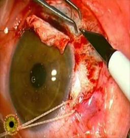
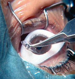
A tear or hole of the retina that leads to a peripheral retinal detachment causes the loss peripheral vision. This situation will progress to a full retinal detachment and loss of all vision if the problem is not repaired. The surgical repair of a retinal detachment is usually successful in reattaching the retina. Scleral buckling is the standard treatment for detached retinas. The surgery is done in anoperating room under general or local anesthesia. The surgeon identifies the holes or tears either through the operating microscope. The hole or tear is then sealed, either with diathermy, a cryoprobe, or a laser. This resulting scar tissue formed around the retinal tear keep it permanently sealed, so that fluid no longer can pass through and behind the retina. A scleral buckle, which is made of silicone, plastic, or sponge, is then sewn to the outer wall of the eye (the sclera). The buckle is like a tight cinch or belt around the eye. This application compresses the eye so that the hole or tear in the retina is pushed against the outer scleral wall of the eye, which has been indented by the buckle. The buckle may be left in place permanently. It usually is not visible because the buckle is located half way around the back of the eye and is covered by the conjunctiva.
About Eye
Eye Diseases
Vision problems
Find Cost
Surgeries & Treatments
Best Eye Hospitals
Testimonials
Get Free Quotes
Top Hospitals In India
Top Hospitals In Turkey
Top Hospitals In Mexico
Top Hospitals In Costa Rica
Top Hospitals In Singapore
Top Hospitals In UAE
Top Hospitals In Australia
Top Hospitals In Malaysia
Top Hospitals In Thailand
Top Hospitals In Phillipines