- cataract surgery
- chalazion surgery
- intraocular lens implant surgery
- lasik surgery
- scleral buckling surgery
- Blepharoplasty Eyelid Surgery Guide
- Cataract Surgery Complete Guide
- Cross-Linking (CXL) for Keratoconus Guide
- DALK/DSEK Corneal Transplant Guide
- Glaucoma Drainage Implant (Ahmed Valve) Guide
- Glaucoma Surgery: Trabeculectomy Guide
- Intacs Corneal Implants for Keratoconus Guide
- LASIK vs. PRK Comparison Guide
- Macular Hole Surgery Complete Guide
- Phakic IOL (ICL) Implantation Guide
- Phakic IOL Implantation (ICL) Guide
- PRK Surgery Guide
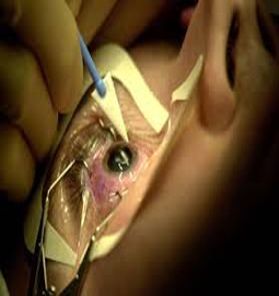
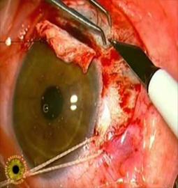
- Pterygium Excision with Graft Surgery Guide
- Ptosis Repair Surgery Guide
- Refractive Lens Exchange Guide
- Retinal Detachment Surgery Guide
- SMILE Laser Eye Surgery Guide
- Strabismus (Squint) Surgery Complete Guide
- Vitrectomy Surgery Complete Guide
- YAG Laser Capsulotomy Guide
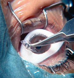
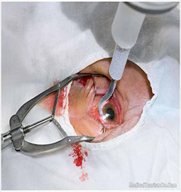
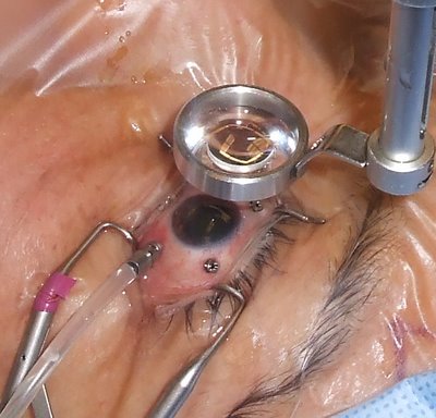
Cataracts occur when the lens of the eye becomes clouded, which results in a decreased vision that gets progressively worse over time. cataracts are a natural process and, thus far, an unavoidable part of aging. Cataract eye surgery involves the removal of the natural, clouded lens of the eye and its subsequent replacement with a clear, artificial lens. Cataract surgery is generally performed with minimal sedation and generally takes less than 30 minutes. Therefore the surgery does not put significant strain on the heart or the lungs. The most common type of cataract eye surgery performed in the U.S. today is an outpatient procedure using a process called phacoemulsification. Cataract surgery alone or combined with trabeculectomy must be considered in the treatment of angle closure glaucoma.
There are two types of cataract surgery:
Phacoemulsification, or phaco.
Extracapsular surgery
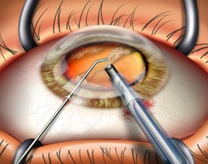
Cataract surgery slightly increases the risk of retinal detachment. A cataract surgery poses risks, such as infection and bleeding. So before cataract surgery, your doctor may ask you to temporarily stop taking certain medications which increases the risk of bleeding during surgery.
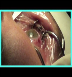
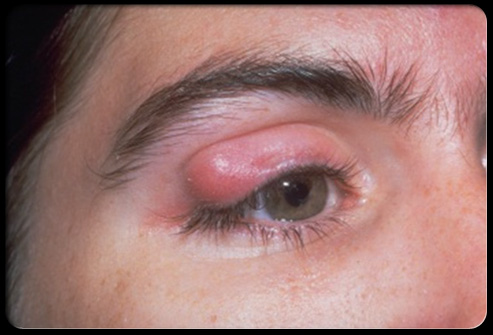
Chalazion is a lump under the eyelid but it is actually a tumor and the lump can reach even greater proportions. Chalazion surgery is a quick, virtually painless procedure performed in less than 15 minutes under a local anaesthetic. It is a minor office surgical procedure. People who is having cyst are often prone to getting more in the future, either at the same areas or other areas of the eyelids
When the chalazion is very small, it can be removed through a small cut at the back of the eyelid. Before the treatment, if you have problems with your blood pressure, your heart or your lungs, everything should be under control. During the operation, local anesthetics is applied with a small injection into the eyelid which makes the area of the operation numb, Now the eyelid is lifted so that the surgeon has access to the back surface of it and a small cut of about 3mm is made just on top of the chalazion and the lump is then removed and pressure is applied for a few minutes to stop any oozing of blood that may occur because of the operation performed. In this procedure, there is no need for stitches and since the cut is at the back of the eyelid, it doesn’t show and the result will be excellent.
Intraocular lenses(IOLs) is the product of choice for both cataract patients and eye surgeons. Intra ocular lenses are made from clinical grade plastics. Intraocular lenses (IOLs) Products with a unique double-convex design, these intraocular lenses combine principles of diffraction, refraction and apodization to produce clear vision along a continual range of distances.
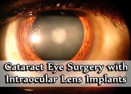
Nonfoldable Intraocular Lens Implant
Until recently, ophthalmologists were restricted to the smallest incision they could create. This was due to the size of the non foldable intraocular lens implant. This type of incision is 5 - 7 mm and may require sutures for wound closure.
Foldable Intraocular Lens Implants
With the advance of foldable IOLs, lenses can be implanted through the same small incision that is created in the phaco procedure. These IOLs are made of a flexible material allowing them to be folded for implantation.
Causes
Cataract is the clouding of the natural lens which is the part of the eye responsible for focusing light and producing clear, sharp images. The lens is contained in a sealed bag or capsule. As old cells die they become trapped within the capsule. Over time, the cells accumulate causing the lens to cloud, making images look blurred or fuzzy. This is a leading cause of visual loss among adults over 55years of age. Eye injuries, certain medications, and diseases such as diabetes and alcoholism have also been known to cause cataracts.
Procedure:
During implant surgery, these lenses are placed in the eye via a very small incision that is actually shorter than the diameter of the multifocal intraocular lens itself. In most cases, intraocular lens surgery can be performed in less than an hour. The surgeon places the ReSTOR IOL in the recipient's eye, where it unfolds itself as it uses built-in anchors to secure itself in its proper position. An intra ocular lens may be placed in the space between the cornea and the iris, be attached to the iris or be placed behind the iris and pupil. Unlike older intraocular lenses, the ReSTOR product is able to move and bend within the eye, producing clearer and more natural vision. Most lens implants are available in a standard power range of +10D to +30D.
These intraocular lenses are available to cataract patients who are otherwise in generally good health. While the surgical procedure used to insert them is one of the safest and most common operations performed.
Risksassociated with it:
The risks include eye infection and inflammation, bleeding, retinal detachment and the possibility of glaucoma. Diabetics and infection-prone individuals should discuss their overall health with a doctor in advance of surgery, as these conditions can raise the risk of unwanted side effects occurring.
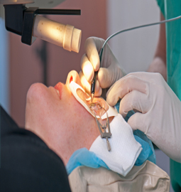
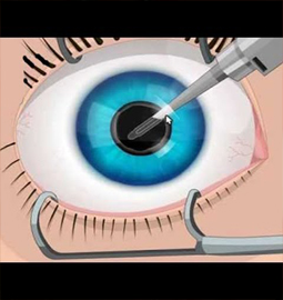
The word LASIK is an acronym for Laser- Assisted In-Situ Keratomileusis (LASIK) and is currently a popular surgical procedure that is being performed by Eye Doctors to improve vision by correcting refractory errors. This procedure utilizes a highly specialized laser which is designed to treat refractive errors, improve vision, and reduce or eliminate the need for glasses or contact lenses.
LASIK eye surgery has become the most popular vision surgery in the country. The surgery improves the vision in patients who have nearsightedness, farsightedness, and astigmatism. LASIK surgery usually takes less than one minute per eye. Treatment involves reshaping the cornea - the protruding portion of surface of the eye - to improve vision.
LASIK Eye Surgery is the procedure of choice and can be elected to be done for a person who is dependent on glasses or contact lens for a prolonged period. It corrects the refractory errors in patients who have -
Myopia or Near-sightedness
Hypermetropia or Far-sightedness
Astigmatism
In order to perform this procedure the following conditions should be satisfied:
The patient needs to be over 18 years and healthy
Should be no infection, glaucoma or dry eyes
Refraction of the eye should be stable for at least a year
Should not be on prescription drugs, like oral prednisone
Not be pregnant or nursing
Not suffering from degenerative or autoimmune diseasePreparation for LASIK Eye Surgery starts well before the surgery. The doctor should be told about the medical history, medications currently being taken and medications allergic to, if any.
Precautions:
If using soft contact lens, the patient must revert to using glasses for at least two weeks prior to the baseline evaluation. This is to restore the normal shape of the cornea. If using hard contact lens the user should stop using them from 6 weeks prior to the surgery. Usage of creams, lotions or cosmetics on the face and eyes should be avoided a day prior to the surgery.
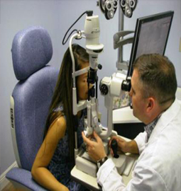
Before accepting LASIK eye surgery, a thorough baseline evaluation of the eye is required by the ophthalmologist. Once the patient's eyes are prepared for surgery a computer and a low wattage laser is used to scan and map the cornea. This information creates a map of the cornea and it helps the computer to determine its exact shape. The surgeon uses this information to calculate the exact amount and depth of corneal tissue to be removed during the operation to get the right shape.
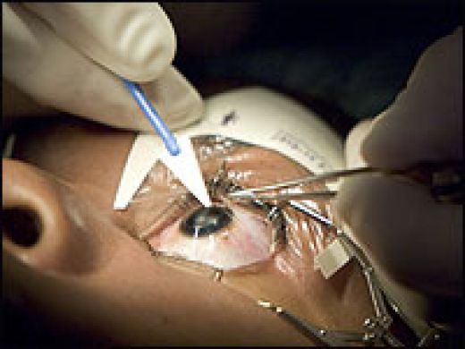
Scleral buckling surgery is the most common way to treat a retinal detachment.In this procedure a piece of silicone or sponge is sewn onto the sclera at the site of a retinal tear which pushes the sclera toward the retinal tear. This buckle supports the retina against the sclera till the scarring seals the tear.
Procedure:
A scleral buckle is a piece of silicone sponge, rubber, or semi-hard plastic that is sewn into place on the outside of the eye. The buckling element is usually left in place permanently. This pushes in, or "buckles," the sclera toward the middle of the eye thus relieving the pull on the retina, allowing the retinal tear to settle against the wall of the eye. The buckle effect may cover only the area behind the detachment, or it may encircle the eyeball like a ring. This does not prevent a retinal break from opening again. Cryopexy, diathermy or laser photocoagulation is used to scar the retina and hold it in place until a seal forms between the retina and the layer beneath. The seal holds the layers of the eye together thus preventing fluid from getting between them. A gas bubble can also be injected into your eye to flatten the retina.
This procedure is done in a hospital as an outpatient procedure. A local or general anesthesia may be used. Both the eyes may be patched to keep the detachment from spreading. Eyedrops mayb be used to have the pupils dilated. The procedure typically takes 1 to 2 hours.
Vitrectomy has been shown to greatly improve visual acuity in many people who have severe vitreous hemorrhage that has not cleared on its own. A vitrectomy can decrease the risk of severe bleeding in people who have begun to have bleeding into the vitreous gel. It can also reduce the risk of severe bleeding in people with growth of abnormal blood vessels in the iris.
Surgery can restore some vision lost as a result of traction retinal detachment and may help prevent further detachment. But the results tend to be better when the detachment has not affected the center of the retina (macula) and the central vision it provides.
A tear or hole of the retina that leads to a peripheral retinal detachment causes the loss peripheral vision. This situation will progress to a full retinal detachment and loss of all vision if the problem is not repaired. The surgical repair of a retinal detachment is usually successful in reattaching the retina. Scleral buckling is the standard treatment for detached retinas. The surgery is done in anoperating room under general or local anesthesia. The surgeon identifies the holes or tears either through the operating microscope. The hole or tear is then sealed, either with diathermy, a cryoprobe, or a laser. This resulting scar tissue formed around the retinal tear keep it permanently sealed, so that fluid no longer can pass through and behind the retina. A scleral buckle, which is made of silicone, plastic, or sponge, is then sewn to the outer wall of the eye (the sclera). The buckle is like a tight cinch or belt around the eye. This application compresses the eye so that the hole or tear in the retina is pushed against the outer scleral wall of the eye, which has been indented by the buckle. The buckle may be left in place permanently. It usually is not visible because the buckle is located half way around the back of the eye and is covered by the conjunctiva.


About Eye
Eye Diseases
Vision problems
Find Cost
Surgeries & Treatments
Best Eye Hospitals
Testimonials
Get Free Quotes
Cataract Surgery
Intacs
Astigmatic Keratotomy(AK)
Canalicular Tear Repair
Chalazion Surgery
Conductive Keratoplasty(CK)
Corneal Transplantation
Lasik Surgery


Top Hospitals In India
Top Hospitals In Turkey
Top Hospitals In Mexico
Top Hospitals In Costa Rica
Top Hospitals In Singapore
Top Hospitals In UAE
Top Hospitals In Australia
Top Hospitals In Malaysia
Top Hospitals In Thailand
Top Hospitals In Phillipines